VISTAL
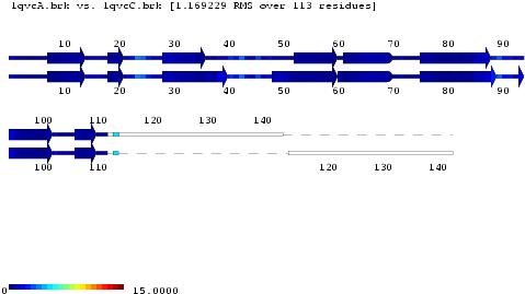
VISTAL is a two-dimensional visualization tool for structural alignments. It describes structures as a series of secondary structure elements, and places matched residues, one on top of each other, colored according to the three-dimensional distance of their Ca atoms.
Download
Click here to download VISTAL.
Example
VISTAL view without wrapping of alignment (multiple images).


We display the images in a table, side by side. Source:
[[Image:ska_mult.1.jpg]]
[[Image:ska_mult.2.jpg]]
[[Image:ska_mult.3.jpg]]
[[Image:ska_mult.4.jpg]]
[[Image:ska_mult.5.jpg]]
VISTAL view with wrapping of alignment.
Reference
Kolodny, R. and Honig, B. (2006) VISTAL – A New 2D Visualization Tool of Protein 3D Structural Alignments. Bioinformatics 22:2166-2167.
Acknowledgments
VISTAL is supported by a funding from the National Institutes of Health Grant # GM30518 and GM074958.
Developed in the Honig Lab
Questions
For questions related to VISTAL, contact honig_software@c2b2.columbia.edu.